Scientific Chronicle
Spring and Children’s Immune Development: Molecular Insights for Enhanced Resilience
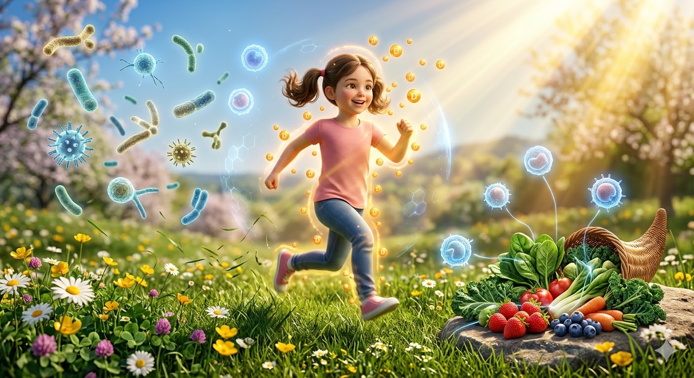
Spring is not just a season of longer days and blooming landscapes—it’s a period that exerts profound biological effects on children’s immune development. After months of limited sunlight and reduced microbial exposure in winter, spring creates a unique window for immune system reinforcement.
Sunlight, Vitamin D, and Immunomodulation
Increased UVB exposure during spring drives cutaneous synthesis of vitamin D3, which is hydroxylated in the liver to 25(OH)D3 and then converted in the kidney to the biologically active calcitriol. Calcitriol binds to the vitamin D receptor (VDR) expressed in thymic epithelial cells, dendritic cells, and T lymphocytes. Activation of VDR modulates transcription of key genes involved in immune regulation, including CAMP (cathelicidin antimicrobial peptide) and DEFB4 (defensin beta 4), enhancing innate defense against bacterial, viral, and fungal pathogens. Additionally, vitamin D signaling suppresses pro-inflammatory cytokines such as IL-6, TNF-α, and IL-17, while promoting regulatory T-cell (FOXP3+ Treg) differentiation through pathways involving STAT5 and TGF-β signaling, contributing to immune tolerance and reduced risk of autoimmunity.
Microbial Exposure and Immune Training
Spring encourages outdoor activity, exposing children to environmental microbes that are critical for shaping immune networks. According to the hygiene hypothesis, limited early-life microbial encounters—common in urban lifestyles—may impair immune education, increasing susceptibility to allergies and autoimmune diseases. Children in farm or rural settings experience higher microbial diversity, which drives expansion of Th1 and Treg populations while balancing Th2 responses, lowering the incidence of asthma, eczema, and allergic rhinitis. Environmental exposure also influences the gut microbiome, where commensals modulate immune development through TLR (Toll-like receptor) signaling and the production of short-chain fatty acids like butyrate, which enhances histone acetylation in Tregs and intestinal epithelial cells, reinforcing mucosal barrier integrity.
Seasonal Nutrition: Molecular Support for Immunity
Spring’s abundance of leafy greens, berries, and prebiotic-rich vegetables supports immune function at the molecular level. Nutrients such as vitamin C, flavonoids, and folate enhance NF-κB regulation, antioxidant responses, and lymphocyte proliferation. Prebiotic fibers promote expansion of Bifidobacterium and Lactobacillus, which modulate IL-10 production, dampening inflammatory cascades. Sulfur-containing compounds in garlic and chives induce Nrf2-mediated antioxidant pathways, further strengthening cellular defenses.
Physical Activity and Lymphatic Activation
Regular outdoor play stimulates muscle contractions that enhance lymphatic circulation, promoting immune surveillance by facilitating trafficking of dendritic cells, naïve T-cells, and NK cells. Moderate activity enhances IFN-γ production by NK cells and cytotoxic T lymphocytes, supporting antiviral defense. Overexertion, however, may transiently elevate cortisol, suppressing IL-2 production and reducing T-cell proliferation, highlighting the importance of balanced activity.
Challenges: Allergens and Seasonal Viruses
Spring also introduces immunological stressors. Pollen exposure activates IgE-mediated mast cell degranulation, while seasonal viruses such as RSV trigger TLR3/7 and RIG-I pathways, leading to type I interferon responses. Strategic mitigation—air filtration, hand hygiene, and vaccination when available—can reduce these immune burdens.
Takeaway for Parents and Practitioners
- Optimize sunlight exposure to support VDR signaling and vitamin D–dependent antimicrobial pathways.
- Encourage outdoor play for microbial diversity and Th1/Treg balance.
- Include seasonal fruits, vegetables, and prebiotics to modulate NF-κB, Nrf2, and gut–immune crosstalk.
- Promote regular moderate physical activity to stimulate lymphatic trafficking and NK/T-cell activation.
- Monitor and manage allergen and viral exposure to prevent immune overactivation.
Conclusion
Spring acts as a natural immunological booster, enhancing both innate and adaptive pathways in children. By integrating sunlight, microbial exposure, nutrition, and physical activity, parents can harness the season’s molecular advantages to strengthen immune resilience, reduce inflammatory risks, and support long-term health.
Organ Banks and Organ Preservation: Importance, Applications, and What Can Be Stored

As a PhD-trained immunologist, I have worked for years at the interface of immune biology, transplantation science, and clinical preservation technologies. Organ banking is not a futuristic concept or a luxury of advanced healthcare systems; it is a medical necessity that directly determines survival, transplant success, and long-term immune tolerance.
From an immunological standpoint, the way an organ or biological tissue is preserved is just as important as donor–recipient matching. Poor preservation alters antigen presentation, increases ischemia–reperfusion injury, and amplifies inflammatory cascades that ultimately raise the risk of rejection.
Why Organ Preservation Matters
Organ preservation is the backbone of modern transplantation medicine. Once an organ is removed from the donor, a biological countdown begins. Cellular hypoxia, oxidative stress, endothelial damage, and immune activation start immediately. Organ banks exist to slow, control, and biologically manage this process.
Effective preservation:
- Maintains cellular viability and structural integrity
- Reduces ischemia–reperfusion injury
- Limits innate immune activation
- Improves graft survival and long-term function
- Expands the usable donor pool
From an immunological perspective, every additional hour of proper preservation reduces antigenic chaos and improves immune adaptation after transplantation.
What Can Be Stored in Organ Banks?
Organ banks are not limited to whole organs. Depending on the bank type and preservation infrastructure, the following biological materials can be safely stored and clinically used:
1. Solid Organs
These are typically preserved under hypothermic or normothermic perfusion systems:
- Kidney
- Liver
- Heart
- Lung
- Pancreas
Kidneys are the most commonly stored organs due to higher tolerance to ischemia, while hearts and lungs require highly controlled preservation conditions.
2. Tissues
Tissue banking is a critical but often underestimated part of transplantation medicine:
- Cornea
- Skin grafts
- Bone and bone marrow matrix
- Tendons and ligaments
- Heart valves
- Blood vessels
These tissues are used not only in transplantation but also in trauma care, reconstructive surgery, burn management, and cardiovascular surgery.
3. Cellular Products
From an immunological and regenerative medicine standpoint, this category is expanding rapidly:
- Hematopoietic stem cells
- Mesenchymal stem cells
- Immune cells for therapy and research
- Isolated pancreatic islet cells
These materials are stored using cryopreservation protocols that maintain cell viability, surface markers, and functional immune properties.
4. Reproductive and Perinatal Biological Materials
Although not classical “organ banking,” these materials play a major role in immune tolerance and regenerative medicine:
- Umbilical cord blood
- Placental tissues
- Amniotic membrane
Cord blood banking, in particular, has strong immunological value due to the naïve immune profile of neonatal stem cells.
Applications of Organ and Tissue Banking
Organ banks support a wide range of clinical and scientific applications:
- Life-saving organ transplantation
- Emergency trauma and burn treatment
- Congenital defect repair
- Cardiovascular surgery
- Immune and stem cell therapies
- Biomedical and immunological research
In research settings, preserved tissues allow controlled investigation of immune responses, rejection mechanisms, and tolerance pathways.
Immunological Challenges in Organ Banking
From my perspective as an immunologist, preservation is not just a technical process; it is an immune-modulating intervention. Improper storage can:
- Increase MHC expression
- Activate complement pathways
- Promote cytokine release
- Prime the graft for rejection before transplantation even occurs
Modern organ banks therefore integrate immunology, biochemistry, and bioengineering to optimize outcomes.
Final Perspective
Organ banking represents the silent infrastructure of modern medicine. Without it, transplantation, regenerative therapies, and advanced surgical care would simply not exist.
As an immunologist, I consider proper organ and tissue preservation not merely a logistical step, but a decisive immunological intervention that shapes patient survival, graft acceptance, and long-term health outcomes.
This is why continued investment in organ banking technologies, regulation, and research is not optional; it is essential.
Pancreas Transplantation in Type 1 and Type 2 Diabetes
Clinical Reality, Immunological Limits, and Human Cost

As a medical immunologist, I approach pancreas transplantation with both scientific respect and clinical caution. For patients with diabetes, especially Type 1 diabetes, this procedure represents one of the few interventions capable of restoring physiological insulin production. At the same time, it introduces a lifelong immunological burden that must never be underestimated.
Indications and Patient Selection
Pancreas transplantation is primarily designed for patients with Type 1 diabetes mellitus who suffer from severe glycemic instability, recurrent hypoglycemia, or progressive complications despite optimal medical management. In selected cases of Type 2 diabetes, transplantation may be considered, but only when beta-cell failure is evident and insulin resistance remains limited. This group is small and requires extremely careful evaluation.
Optimal Age for Transplantation
From an immunological and surgical standpoint, the most suitable candidates are typically between 18 and 50 years of age. Younger patients generally demonstrate better vascular integrity, fewer comorbidities, and more predictable immune responses. In older individuals, chronic inflammation, cardiovascular disease, and immune dysregulation significantly increase the risk of complications and graft failure.
Benefits and Potential Advantages
The primary benefit of pancreas transplantation is the restoration of endogenous, glucose-responsive insulin secretion. Successful transplantation can eliminate the need for exogenous insulin, stabilize blood glucose levels, and reduce episodes of severe hypoglycemia. Over time, sustained normoglycemia may slow the progression of diabetic microvascular damage, improving long-term outcomes.
Risks, Limitations, and Immunological Cost
This procedure carries substantial risks. Surgical complications, graft thrombosis, acute rejection, and infection remain significant concerns. More importantly, lifelong immunosuppressive therapy is unavoidable. From an immunological perspective, this represents a permanent alteration of immune balance, increasing vulnerability to infections, malignancies, and systemic inflammatory disturbances. The immune system is controlled, not neutralized, and this distinction matters.
Pancreas Transplantation in Children
In pediatric patients, pancreas transplantation should be considered only in exceptional circumstances. Most children with Type 1 diabetes achieve acceptable control with modern insulin delivery systems and continuous glucose monitoring. In rare cases of uncontrollable hypoglycemia or extreme metabolic instability, transplantation may be discussed. However, the psychological burden, long-term medication dependency, and impact on immune development demand extreme caution.
Final Perspective
Pancreas transplantation is not a cure for diabetes. It is a complex therapeutic substitution that trades metabolic control for immunological compromise. When applied to carefully selected patients, the benefits can be life-changing. When applied indiscriminately, the consequences can be severe.
As a medical immunologist, I believe this intervention must remain a highly specialized option, guided by evidence, ethics, and a clear understanding of immune tolerance. Advances in diabetes care continue to evolve, and transplantation should be reserved for cases where its benefits clearly outweigh its long-term immunological costs.
Immunoendocrine Mechanisms Underlying Risky and Self-Destructive Behavior
As an immunologist, I am increasingly intrigued by the profound ways in which the immune and endocrine systems jointly shape behavior—a field we term immunoendocrinology. Recent research indicates that sex hormones, stress mediators, and immune signaling molecules collectively influence risk perception, social behavior, and decision-making in both humans and animals.
Animal Models of Immunoendocrine Behavior
In certain animal species, behaviors that appear “self-destructive” are in fact tightly regulated by immunoendocrine mechanisms. Consider BobaK marmots: these animals sometimes engage in unusually affiliative behavior toward wolves, their natural predators. At first glance, such actions seem evolutionarily maladaptive. However, studies suggest that fluctuations in testosterone and estrogen modulate neural circuits responsible for social dominance, risk assessment, and fear responses.
Simultaneously, cytokines and chemokines produced in response to environmental stressors convey information from the immune system to the central nervous system, influencing exploratory and risk-prone behaviors. These interactions can transiently bias an animal toward seemingly irrational choices, such as approaching a predator.
Parallel Phenomena Across Species
A parallel phenomenon is observed in juvenile rabbits. When experiencing maternal separation or social conflict, they occasionally move toward predator dens—behavior that, while life-threatening, reflects an interplay between stress hormones (cortisol and catecholamines), sex hormones, and immune signaling. Cytokine-mediated modulation of amygdala and prefrontal circuits can transiently suppress fear responses, promoting high-risk exploratory behavior. This suggests that what appears as “irrational” behavior is, in fact, an evolutionarily conserved response orchestrated by the immunoendocrine axis.
Human Implications
Humans, too, are not exempt from these mechanisms. For instance, decision-making under emotional stress—such as impulsively confronting a high-risk social situation or engaging in potentially harmful financial choices—can be linked to fluctuations in sex hormones, cortisol, and immune mediators. Inflammatory cytokines, for example, can affect neural circuits involved in reward processing and risk evaluation, subtly predisposing individuals to decisions they might later regret. In this context, so-called “stupid” choices are not purely cognitive errors but may reflect an underlying biological state shaped by the immune and endocrine systems.
Conclusion
Understanding the immunoendocrine basis of risky decision-making illuminates behaviors that were traditionally interpreted as irrational. It provides a mechanistic framework linking hormones, immune signaling, and neural circuits to both adaptive and maladaptive behaviors, across species. By studying these conserved pathways, we gain insights not only into the ecology of animal behavior but also into the biological underpinnings of human social, emotional, and cognitive decisions.
The Impact of Thalassemia on Hair and Beard Growth — Scientific Explanation, Causes, Diagnosis, and Treatment Options
Abstract
Thalassemia (particularly transfusion-dependent beta-thalassemia) can affect hair and beard health in several ways, ranging from diffuse hair loss and thinning to irregular or weak beard growth. These effects result from a combination of chronic anemia, iron overload, endocrine complications secondary to iron deposition, nutritional deficiencies, and, in some cases, side effects of treatment. Addressing the underlying cause along with topical/systemic therapies and, in selected cases, hair transplantation under specific medical conditions can be beneficial. (References and details provided in the text.)
PMC+1
Brief Overview: Types of Thalassemia
- Alpha-thalassemia: Deletions or mutations in alpha-globin genes — spectrum ranges from silent carriers (trait) to hydrops fetalis.
- Beta-thalassemia: Includes major (transfusion-dependent / TDT), intermedia, and minor (carrier).
- Clinical relevance: The severity of the disease (e.g., frequency of transfusions) determines the risk of secondary complications such as iron overload and subsequent endocrine/systemic effects.
NCBI+1
Why Can Thalassemia Cause Hair Loss or Poor Beard Growth? (Mechanisms)
- Chronic anemia and metabolic stress: Reduced peripheral oxygenation may slow down hair growth and shift follicles into the telogen (resting) phase.
- Iron overload: Repeated transfusions lead to iron deposition in tissues, causing cellular damage and oxidative stress — these mechanisms can impair the skin, hair follicles, and endocrine-hormonal axes.
- Endocrine disorders: Iron deposition in the pituitary and gonadal glands can lead to hypogonadism or thyroid dysfunction, both linked to hair loss and irregular beard/hair growth. Studies show high prevalence of sexual dysfunction, delayed puberty, and related endocrine abnormalities in TDT patients.
- Nutritional and metabolic deficiencies: Zinc, vitamin D, protein, or other micronutrient deficiencies may coexist and impair hair growth.
- Drug or treatment-related effects: Certain chelation therapies or chronic infections may contribute.
- Chronic inflammation and oxidative stress: Persistent inflammatory states can disrupt follicular cycling.
Summary: The dominant pathway is iron overload → endocrine damage → hormonal/nutritional imbalance → hair loss/weak growth.
MDPI+1
Common Clinical Manifestations in Thalassemia Patients (Hair-Related)
- Diffuse hair thinning, especially during adolescence and adulthood in patients with high iron load.
- Patchy or thin beard growth in males (often due to hypogonadism or low testosterone).
- Alopecia patterns secondary to nutritional or drug-induced causes.
Accurate diagnosis requires dermatological examination, evaluation of hair loss patterns, and laboratory investigations.
PMC
Medical Evaluation (Recommended Laboratory Tests)
- Complete blood count (CBC) – to assess anemia.
- Ferritin, TIBC, NTBI/LIC – to evaluate iron overload (ferritin alone is not sufficient but is a first-line marker).
- Endocrine panel: FSH, LH, testosterone (in males), TSH, Free T4, and diabetes profile if indicated.
- Micronutrient assessment: Zinc, vitamin D, and serum iron (may vary in carriers).
- Specialized evaluations: Hematology consultation and, when needed, liver/heart MRI for iron concentration (LIC, cardiac MRI T2*).
Identifying and treating the underlying cause (e.g., treatable hypogonadism) is the key step, as many solutions stem from there.
General and Specific Treatments for Hair/Beard Problems in Thalassemia
A) Etiology-focused treatment (top priority)
- Iron overload management: Optimize transfusion schedules and chelation therapy (deferasirox, desferrioxamine, etc.) to prevent or slow endocrine complications. Controlling iron overload is essential to reduce secondary damage.
- Endocrine therapy: In confirmed hypogonadism, hormone replacement (e.g., testosterone in males or thyroid hormone management) can help restore hair and beard growth. Requires close cooperation between hematology and endocrinology.
B) Nutritional and supportive therapy
- Correct deficiencies in zinc, vitamin D, and other micronutrients. (Iron supplementation should be avoided unless medically justified, as iron overload is common in thalassemia.)
- Maintain a protein-rich diet and consider supplementation as needed.
C) Topical/pharmacologic treatments for hair growth
- Topical minoxidil: Effective for diffuse thinning (though does not address the underlying cause).
- Finasteride: In androgenic alopecia, for men only and with caution after full assessment.
- Scalp care: Gentle washing and avoidance of mechanical stress.
D) Surgical/interventional treatments (Hair transplantation)
Detailed in the next section.
Hair Transplantation in Thalassemia — Risks, Benefits, and Safety Criteria
Is thalassemia a contraindication to hair transplantation?
Generally, thalassemia is not an absolute contraindication, but thorough evaluation by a hematologist and hair transplant surgeon is mandatory.
Potential issues to assess:
- Hemoglobin level and anesthesia tolerance: Severe anemia may increase anesthesia risk — Hb should be optimized preoperatively.
- Bleeding or coagulation disorders: Platelet count and coagulation profile must be checked.
- Infection and wound healing: Chronic infections or iron overload may impair healing; infection control is essential.
- Endocrine status: Untreated hypogonadism or hormonal disorders may limit cosmetic results.
- Chelation or other medications: Review possible drug–anesthetic/antibiotic interactions.
- Donor site healing: Must be evaluated in patients with poor regenerative capacity.
Benefits (for well-selected patients):
- Restoration of hair/beard density where viable donor follicles exist.
- Improved self-esteem and quality of life — only after systemic stability and safety optimization.
Pre-transplant checklist:
- Hematology clearance (Hb, platelets, coagulation, ferritin/LIC, transfusion and chelation plan).
- Endocrine evaluation (thyroid and sex hormones).
- Infection control (skin/systemic).
- Joint planning between surgeon and hematologist; transfusion or medication adjustment may be needed pre-op.
Dermatoloji Dergisi+1
Emerging and Advanced Therapies Indirectly Improving Hair Health
Recent advances in stem cell and gene therapy for beta-thalassemia have shown promising results, reducing transfusion dependency and long-term iron overload, thereby indirectly improving hair and beard health.
Examples include exa-cel / Casgevy (exagamglogene autotemcel), FDA-approved in January 2024 for transfusion-dependent beta-thalassemia, and beti-cel / Zynteglo from earlier generations — both of which significantly reduce iron overload and endocrine complications over time.
hematology.org+2, Çocuk Hastanesi+2
Practical Recommendations — Simplified Algorithm for Physicians/Patients
- Comprehensive clinical evaluation (hematology, endocrinology, nutrition).
- If iron overload or suboptimal chelation: improve management or refer to a specialized center. TIF
- If hypogonadism/hormonal deficiency: initiate hormone replacement under endocrinologist supervision.
- Nutritional support and topical/scalp therapy (minoxidil, care routine).
- For hair transplant candidates: perform full risk–benefit assessment and coordinate timing between hematology and surgery teams.
Dermatoloji Dergisi
Notes for Immunology Readers (and SEO Optimization)
- As an immunology expert, emphasize the relationship between oxidative stress, systemic inflammation, and follicular health — this mechanistic insight strengthens the scientific depth and SEO relevance of the article.
- Highlight the multidisciplinary approach (hematology, endocrinology, dermatology, reconstructive surgery) in management.
Nature
Key References (For Further Reading)
- De Sanctis V. Growth and endocrine disorders in thalassemia. (Comprehensive review on endocrine dysfunctions in thalassemia). PMC
- Evangelidis P. Endocrinopathies in Hemoglobinopathies (2023 review on iron overload and oxidative stress). MDPI
- Thalassemia International Federation (TIF) Guidelines for TDT management.
- Casgevy / Exagamglogene Autotemcel gene therapy updates and FDA approval (Jan 2024). hematology.org+1
- Clinical Reviews (2024): Pathophysiology and novel therapies in beta-thalassemia. MDPI
Why Mineral Carbonated Water Should Be Avoided After Blood Donation
And Whether Sweet Drinks Are Harmful or Helpful for Diabetic Donors

Blood donation is a safe and life-saving procedure when followed by proper post-donation care. However, inappropriate fluid or carbohydrate intake immediately after donation can lead to avoidable complications, especially in individuals with metabolic disorders such as diabetes. As an immunologist, I will clarify several common misconceptions.
Why Carbonated Mineral Water Is Not Recommended After Blood Donation
After donating blood, the body experiences a temporary reduction in circulating blood volume. Rapid compensation depends on adequate plasma volume restoration, electrolyte balance, and vascular stability.
Carbonated mineral water, particularly those rich in bicarbonate and dissolved CO₂, is not ideal immediately after donation for several physiological reasons:
1. Gastrointestinal Vasodilation and Discomfort
Carbon dioxide increases gastric distension, which can activate vagal responses and worsen post-donation dizziness or nausea.
2. Transient Acid–Base Shifts
In individuals sensitive to acid–base changes, carbonated beverages may interfere with rapid physiological compensation after mild hypovolemia.
3. Delayed Effective Hydration
Although mineral water contains electrolytes, carbonation slows gastric emptying compared to still water, delaying optimal plasma refilling.
For these reasons, still water, oral rehydration solutions, or non-carbonated isotonic fluids are preferred during the first hours after donation.
Are Sweet Drinks Dangerous for Diabetic Blood Donors?
This question is often misunderstood.
Sweet drinks are not automatically dangerous, and in some cases, they are beneficial, provided they are used correctly and selectively.
In Non-Diabetic Donors
After blood donation, mild transient hypoglycemia may occur, especially in first-time donors or individuals with low body mass. In these cases:
- A small amount of glucose-containing fluid can help stabilize blood pressure and prevent vasovagal reactions.
In Diabetic Donors
For diabetic individuals, the situation requires precision rather than fear.
- Controlled carbohydrate intake is not harmful
- Uncontrolled intake is unnecessary and inappropriate
If a diabetic donor experiences:
- Lightheadedness
- Tremor
- Sweating
- Symptoms suggestive of hypoglycemia
Then a measured amount of juice or glucose solution is clinically appropriate, under supervision.
What should be avoided is:
- Large volumes of high-sugar beverages
- Repeated intake without glucose monitoring
In well-controlled diabetic patients, small, deliberate carbohydrate administration is often protective rather than dangerous.
Which Blood Components Can Be Stored in Blood Banks?
Modern blood banking relies on separating donated whole blood into specific components, each with distinct storage conditions:
1. Red Blood Cells (Packed RBCs)
- Stored at 1–6°C
- Shelf life: up to 42 days
- Used for anemia, trauma, and surgical patients
2. Plasma (Fresh Frozen Plasma)
- Stored at −18°C or lower
- Contains clotting factors and immune proteins
- Used in coagulation disorders and massive transfusions
3. Platelets
- Stored at 20–24°C with continuous agitation
- Shelf life: 5–7 days
- Critical for oncology and hematology patients
4. Cryoprecipitate
- Derived from plasma
- Rich in fibrinogen and factor VIII
- Used in specific bleeding disorders
Each component is stored separately to maximize therapeutic efficacy and immunological safety.
Final Medical Perspective
Post-donation care is not a trivial matter. What a donor consumes immediately after giving blood can influence vascular stability, immune balance, and metabolic safety.
- Carbonated mineral water is not dangerous, but it is physiologically suboptimal immediately after donation.
- Sweet drinks are not inherently harmful, even for diabetic patients, when used strategically and medically.
- Blood donation remains a highly controlled, immunologically safe process when followed by evidence-based care.
As both a scientist and clinician in immunology, I emphasize that precision matters more than assumptions.
Today’s Articles
- Spring and Children’s Immune Development
- Organ Banks and Organ Preservation
- Pancreas Transplantation in Diabetes
- Immunoendocrine Mechanisms in Risky Behavior
- Thalassemia Impact on Hair Loss & Beard Growth
- Mineral Water After Blood Donation
Recent Issues
- March 16, 2026
- March 9, 2026
- March 2, 2026
- February 24, 2026
- February 17, 2026